Citation: News of Beam Diagnostics Belarus 1999 2: 25-26.
MRI features of shoulder rotator calf in healthy subjects.
Goncharov V. V., Divakov M. G., Marchuk V. P., Askerko E. A.
Vitebsk Medical Institute, Vitebsk Diagnostic Center, Vitebsk.
|
|
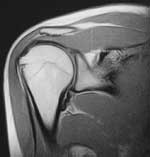
Figure 1. Coronal MR image of the right humeral joint. Coronal
coefficient equal to 0, 989. Sub-acromial space wide equal to 0, 81
sm. (normal values).
1 - supra-spinatous muscle,
2 - deltoid muscle,
3 - humeral joint caput,
4 - scapular acromial outgrowth,
5 - scapular joint socket. |
|
|
|
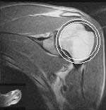
| Figure 2. Coronal MR image of the left humeral joint. Rotation
centres of concave and convex humeral joint components coincides with radius
difference of 0, 66 sm. |
|
|
|
| Figure 3. Axial MR image of the left humeral joint. Supra-spinatous
muscle have a shape of narrowing to distal end parallelogram. |
|