Citation: News of Beam Diagnostics Belarus 1999 2: 33-34.
The case of the necrotic lung dissemination.
Khoruzhik S. A.
Grodno Regional Clinical Hospital, Grodno.
|
|
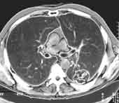
| Figure 1. Chest CT scan shows peripheral mass with cavity in
S6 from the left. Additionally, small number micronodules were hardly seen
in both lungs. Patient refused any future evaluation. |
|
|
|
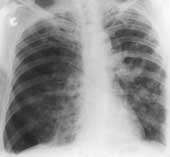
| Figure 2. 7 months later patient was admitted to the hospital
with complaints on right chest pain and suddenly developed shortness of
breath. (а) PA chest radiograph apparently showed disseminated densities
at the both lungs as well as right lung pneumothorax with mediastinum shift
to the left. |
|
|
|
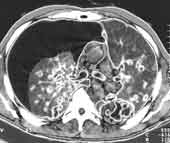
| Figure 2. (b) CT scan shows enlargement of the mass at
the left lung compared to first CT exam as well as multiple nodules with
central cavities even in smallest of them, and right sided pneumothorax.
Not enlarged lymph nodes were found. Left lung mass biopsy verified squamous
cell lung carcinoma. |
|