Citation: News of Beam Diagnostics Belarus 1999 3: 24-26.
MRI presentations of shoulder joint in cases of rotator
calf pathology.
Divakov M. G.1, Askerko E. A.1, Goncharov V.
V.2, Marchuk V. P.2
1Vitebsk Medical Institute, 2Vitebsk Diagnostic
Center, Vitebsk.
|
|
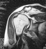
| Figure 1. Oblique coronal MR image (T2w, 96/3091) shows supra-spinatous
muscle lack (arrow). Sub-acromial space narrowed up to 0,42 sm. with humeral
joint decentration. Decentration means not-coincidence of rotation centres
of concave and convex humeral joint components. |
|
|
|
Figure 2. Oblique coronal MR image of the right humeral joint
(PD, 16/3246) shows supra-spinatous muscle defect (arrow). Coronal
coefficient (CC) is 0,71. CC calculated by maximal supra-spinatous
muscle belly width division on its tendinous part length. Normal CC value
was 1,0 +/- 0, 1 in our previous study.
1 - supra-spinatous muscle,
2 - deltoid muscle,
3 - humeral joint caput,
4 - scapular acromial outgrowth,
5 - scapular joint socket. |
|
|
|
| Figure 3. Oblique coronal MR image of the left humeral joint
(T2w, 96/3000) shows supra-spinatous muscle tendon injury (arrow). Marks
- see Figure 2. |
|
|
|
| Figure 4. Oblique coronal MR image of the left humeral joint
(PD, 16/3000) shows supra-spinatous muscle tendon injury with supra-spinatous
muscle superior margin goffering (arrow). Marks - see Figure 2. |
|
|
|
| Figure 5. Oblique coronal MR image of the right humeral joint
(T2w, 96/4018) shows humeral rotator cuff injury with large protuberance
fragment and humeral bone defect of 0,87 sm. depth. |
|