Citation: News of Beam Diagnostics Belarus 1999 3: 28-29.
Percutaneous intrahepatic holangiography.
Chizh G. V.1, Karpovich D. I.2
1Belarussian Medical Academy of Postgraduate Education, 2Minsk
Regional Hospital, Minsk.
|
|
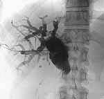
| Figure 1. Percutaneous intrahepatic holangiogram shows dilated
bile ducts with common bile duct sudden narrowing at the level of stone. |
|
|
|
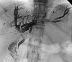
| Figure 2. Multiple filing defects (stones) in moderately dilated
intrahepatic bile ducts. |
|
|
|
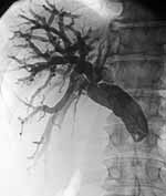
| Figure 3. Intra- and extrahepatic bile ducts dilatation up to
Vater level were common bile duct pipe-like narrowing seen. Thise findings
are characteristic for Vater tumour. |
|
|
|
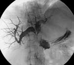
| Figure 4. Patient with pancreatic head carcinoma was underwent
palliative operation of holangioduadenostomy. Follow-up holangiogram shows
partial anastomosis obstruction. |
|