Citation: News of Beam Diagnostics Belarus 1998 4: 18-19.
Purulent meningitis sonography diagnostics in children
of the first year of life.
Hromova T. N.1, Ulezko E. A.2
1Children Regional Hospital, Brest. 27th City
Clinical Hospital, Minsk.
|
|
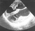
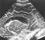
| Figure 1. Clebsielae meningitis. Brain sonogram shows ventricles
widening. |
|
|
|
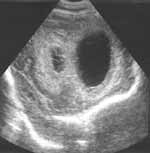
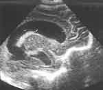
| Figure 2. Clebsielae meningitis. Ventriculitis. |
|
|
|
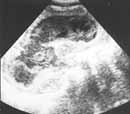
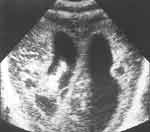
| Figure 3. Clebsielae meningitis. (a) Brain tissue destruction
in acute period. |
|
|
|
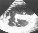
| Figure 3. Clebsielae meningitis. (b) Stage of hydrocephalus
formation. |
|
|
|
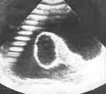
| Figure 4. Clebsielae meningitis. Purulent deposits present in
anterior horn (arrows). |
|
|
|
| Figure 5. Vascular channels changes in staphylococcal meningitis
(arrows). |
|
|
|
| Figure 6. Brain abscess in staphylococcal meningitis (arrows). |
|
|
|
| Figure 7. Staphylococcal meningitis. Sclerotic cyst in thalamus
after abscess. |
|