News of Beam Diagnostics Belarus
Figures Only
Table of Contents Full Text in Russian
 
 
Other Articles:
Previous article
Next article
 

Citation: News of Beam Diagnostics Belarus 1998 4: 20-21.

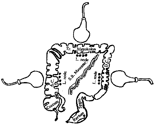
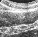
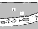
Colonic mesentery sonography examination.

Kushnerov A. I.

Belarussian Scientific Research Institute of Ecological and Professional Pathology, Mogilev.
 
Figure 1. Schematic drawing of probe positions.
 

[View larger version]
Figure 2. Tuberculose lymphadenitis of sigmoid colon. (a) Sonogram.
1 - enlarged mesenteric lymph node,  
2 - sigma lumen,  
3 - hyperechoic regions in mesentery as signs of mesentery inflammation,  
4 - mesenteric edge of sigma,  
5 - edge of sigma mesentery.
 

[View larger version]
Figure 2. Tuberculose lymphadenitis of sigmoid colon. (b) Schema of sonogram.
1 - enlarged mesenteric lymph node,  
2 - sigma lumen,  
3 - hyperechoic regions in mesentery as signs of mesentery inflammation,  
4 - mesenteric edge of sigma,  
5 - edge of sigma mesentery.
Top
To contact Editor of this Internet site please use Feedback Form
Copyright © 2001-2004 by Dr. Serguey Khoruzhik