Citation: News of Beam Diagnostics Belarus 1999 1: 32-34.
Nuclear medicine diagnostics in oncology.
Medvedskij B. E., Grenkov G. I., Tsherbinin Ju. I., Pavlova L. A.
Vitebsk Medical Institute, Vitebsk.
|
|
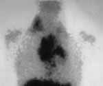
| Figure 1. Colloid Tc-99m hepatic scintigraphy in three projections
shows filing defect in the right lobe. |
|
|
|
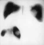
| Figure 2. Ga citrate scintigraphy in Hodgkin lymphoma. (a)
Before treatment increased isotope uptake in mediastinal and right supraclavicle
lymph nodes seen. |
|
|
|
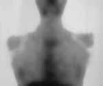
| Figure 2. Ga citrate scintigraphy in Hodgkin lymphoma. (b)
After treatment hot spots are not observed. |
|
|
|
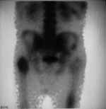
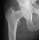
| Figure 4. Bone matastases seen earlier on scintigraphy. (a)
Increased uptake at the upper third of the right and middle third of left
femoral bones. |
|
|
|
| Figure 4. Bone matastases seen earlier on scintigraphy. (b)
Bone radiograph at the same level not revealed metastases. |
|