Citation: News of Beam Diagnostics Belarus 1999 1: 28-30.
Roentgenoscopy + endoscopy = ERHPG.
Doroshko M. V.
Joint venture "Glans", Minsk.
|
|
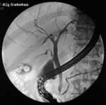
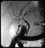
| Figure 1. Endoscopy retrograde holangyopancreatogram shows gallbladder
stones and normal bile ducts tree. |
|
|
|
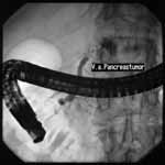
| Figure 2. Stop contrast symptom while trying to fill pancreatic
duct with contrast in patient with pancreatic body carcinoma. |
|
|
|
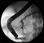
| Figure 3. Chronic pancreatitis signs: pancreatic main duct and
ducts of 2-3rd order dilatation, uneven ducts walls. |
|
|
|
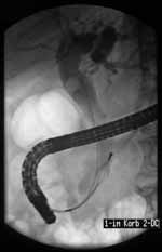
| Figure 4. Dilated holedoh contains stone. Stone extraction procedure
with Dormia FG-22Q basket underway. |
|
|
|
| Figure 5. Idiopathic sclerousing hollangitis. |
|
|
|
| Figure 6. Big cyst of pancreatic head. Cyst internal draining
procedure with use of ТJF-30 duodenoscope underway. |
|