Citation: News of Beam Diagnostics Belarus 1999 1: 21-23.
Recurrent rectal cancer imaging.
Khoruzhik S. A.1,
Fomin K. A.2
1Grodno Regional Clinical Hospital, 2Grodno Medical
Institute, Grodno.
|
|
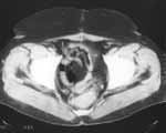
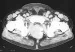
| Figure 1. Patient with rectum cancer have been examined with
CT 5 months after operation to check for recurrence. No recurrence signs
were found. Uterus backward displacement. |
|
|
|
| Figure 2. Radiograph of the left lung. Peripheral lung cancer
developed on the background of the scar seen. |
|
|
|
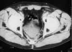
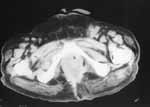
| Figure 3. 11 months after operation bulky recurrent mass between
sacrum and bladder seen. Seminal vesicles are not differentiated. Intratumoral
necrosis seen as region of decreased density inside tumour. |
|
|
|
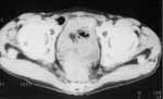
| Figure 4. Recurrent tumor with bladder involvement. |
|
|
|
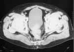
| Figure 5. Recurrent inhomogeneous mass contacting right obturator
internus muscle. Calcificates along left tumor contour as well as inguinal
lymph nodes enlargement from the both sides may be seen. |
|
|
|
| Figure 6. Pubic bone metastasis (arrows) seen as an area of
bone destruction with soft tissue component. Enlarged left inguinal lymph
nodes have not strict contour as sign of extracapsular invasion. |
|

|
 |
Please address your inquiries and comments
about journal and Web site to Serguey
Khoruzhik |
|
Copyright © 2001-2004 by Dr. Serguey Khoruzhik |
|
|
|