Citation: News of Beam Diagnostics Belarus 1999 1: 18-20.
Difficulties and mistakes in beam diagnostics of brain
neoplasias.
Antonenko A. I.
5th City Hospital and Republican Institute of Neurosurgery, Minsk.
|
|
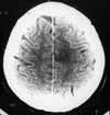
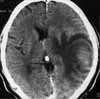
| Figure 1. Left parietal lobe glioblastoma. |
|
|
|
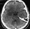
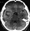
| Figure 2. Viral encephalitis in right temporal lobe. Compare
with Figure 1. |
|
|
|
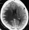
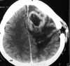
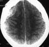
| Figure 3. In right temporoparietal region extensive area of
decreased density seen not enhancing after IV contrast administration. |
|
|
|
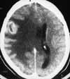
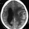
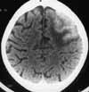
| Figure 4. The same patient as on Figure 3. Follow-up CT scan
in a one month shows metastasis. |
|
|
|
| Figure 5. CT findings of voluminous process without strict border
but with extensive oedema were reported to be glial tumour of the left
temporal lobe. “Grey colour knobby tumour” was totally resected on operation.
Postoperative pathology examination revealed brain abscess. |
|
|
|
| Figure 6. CT scan shows roundish inhomogeneous mass in the right
temporal lobe. Despite some doubts because by previous case (Figure
5), tumour was reported what have been confirmed pathologically. |
|
|
|
| Figure 7. CT scan 1 month after brain haematoma drainage operation.
Two confluent rings like structures at the left frontal lobe were reported
to be abscesses. Lung cancer metastases were diagnosed in this patient
finally. |
|
|
|
| Figure 8. Patient with slowly progressing during for 7 months
neurological deficiency was diagnosed with glial tumour based on CT and
clinical findings. Multiple temporal lobe abscesses were found on operation.
Compare with Figure 7. |
|
|
|
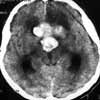
| Figure 9. Pituitary adenoma haemorrhage. |
|
|
|
| Figure 10. In patient with recent brain trauma history CT scan
revealed lens-like isodens collections at the right temporo-parietal region
what was described as subacute epidural haematomas. Pathologic diagnosis
– plasmacytoma. |
|
|
|
| Figure 11. Tuberculome of the left hemisphere. |
|