Citation: News of Beam Diagnostics Belarus 2001 1-2: 34-36.
Present state and perspectives of endovascular interventional
radiology in oncology.
Dudarev V. S., Akinfeev V. V., Vashkevich L. B., Zholnerovich E.
M.
Scientific Research Institute of Oncology and Medical Radiology named
for N. N. Aleksandrov, Minsk.
|
|
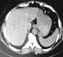
| Figure 1. Hepatocellular carcinoma. (a) Abdominal CT
scan before hemoembolisation shows ill-defined tumour at the 2nd hepatic
segment measuring 2 sm. in diameter. |
|
|
|
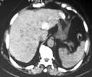
| Figure 1. Hepatocellular carcinoma. (b) After injection
of oil emboliser lesion density increased. |
|
|
|
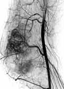
| Figure 2. Osteosarcoma of the lower femur. (a) Angiogram
of the right popliteal artery before arterial hemoembolisation. |
|
|
|
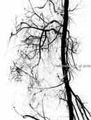
| Figure 2. Osteosarcoma of the lower femur. (b) After
hemoembolisation tumour vessels are not visualised. |
|
|
|
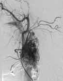
| Figure 3. Left carotid neck paraganglioma. (a) External
carotid angiogram before embolisation. |
|
|
|
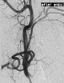
| Figure 3. Left carotid neck paraganglioma. (b) Angiogram
after embolisation. |
|