Citation: News of Beam Diagnostics Belarus 1998 5: 18-20.
Sonographic location of central venous catheter.
Bolbas A. S., Gromyko G. M., Novikov D. V., Karpelev G. M.
Belarussian Scientific Research Institute of Ecological and Professional
Pathology, Mogilev.
|
|
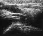
Figure 1. Normal subclavian catheter position. (a) Sonogram
of subclavian region:
1. internal jugular vein,
2. subclavian vein,
3. catheter,
4. shadow from conjunction of sternum and clavicle. |
|
|
|
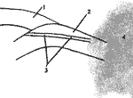
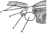
Figure 1. Normal subclavian catheter position. (b) Schema
of sonogram:
1. internal jugular vein,
2. subclavian vein,
3. catheter,
4. shadow from conjunction of sternum and clavicle. |
|
|
|
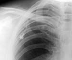
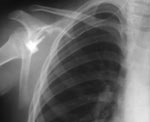
| Figure 1. Normal subclavian catheter position. (c) Radiograph. |
|
|
|
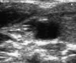
Figure 2. Athypical subclavian catheter position in right jugular
vein. (а) Sonogram:
1. superficial neck muscles,
2. internal jugular vein,
3. catheter,
4. internal carotid artery,
5. right thyroid lobe. |
|
|
|
Figure 2. Athypical subclavian catheter position in right jugular
vein. (б) Schema of sonogram:
1. superficial neck muscles,
2. internal jugular vein,
3. catheter,
4. internal carotid artery,
5. right thyroid lobe. |
|
|
|
| Figure 2. Athypical subclavian catheter position in right jugular
vein. (c) Radiograph. |
|