Citation: News of Beam Diagnostics Belarus 1998 4: 4-6.
Unspecific ulcerative colitis.
Galkin L P., Davidovich T. V.
Homel Medical Institute, Homel.
|
|
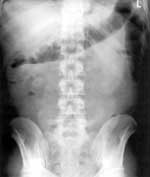
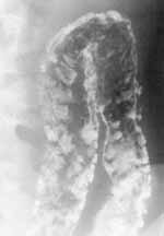
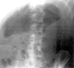
| Figure 1. Non-specific ulcerative colitis. Plain abdomen radiograph
in toxic colon shows redundant amount of gas in the dilated colon. |
|
|
|
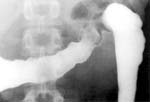
| Figure 2. Early stage non-specific ulcerative colitis. Barium
enema radiograph shows lack of tonus and folds straitening. |
|
|
|
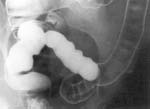
| Figure 3. On the next stage small barium spots on mucous may
be seen. |
|
|
|
| Figure 4. Future non-specific ulcerative colitis progression
results in cobblestoning. |
|
|
|
| Figure 5. Ulcers of different size filled with barium. |
|
|
|
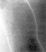
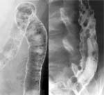
| Figure 6. With the course of time colon wall contours progressively
became roughly wavy. |
|
|
|
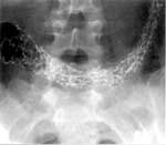
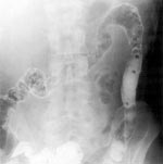
| Figure 7. Barium enema radiograph in non-specific ulcerative
colitis shows pseuopolyps – multiple roundish filling defects of different
size. |
|
|
|
| Figure 8. In chronic stage affected parts may look as atonic
segments with distorted shape, without normal folds and mucous. |
|
|
|
| Figure 9. Non-specific ulcerative colitis complicated with toxic
mega colon. |
|