Citation: News of Beam Diagnostics Belarus 1998 4: 34-37.
Digital subtraction angiography.
Gonchar A. A.1, Gonchar I. A.2
15th City Hospital, 2Scientific Research Institute
of Neurology, Neurosurgery, and Physiotherapy, Minsk.
|
|
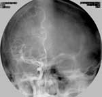
| Figure 1. Conventional frontal angiogram in arterial phase shows
arteriovenous malformation in right temporal region. Aneurysm fills from
anterior and middle brain arteries. |
|
|
|
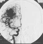
| Figure 2. DSA of the same patient, frontal projection, arterial
phase. Vascular pattern demonstrated in better advantage despite the less
contrast volume was used. |
|
|
|
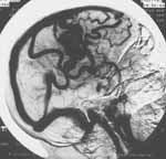
| Figure 3. Right lateral venous phase DSA of the same patient
shows arteriovenous malformation in right temporal region along with multiple
draining veins. |
|