Citation: News of Beam Diagnostics Belarus 1999 1: 4-6.
Roentgenological signs of abdomen pathology on chest
radiographs.
Filippovich N. S.1, Koretko M.V.2
1Belarussian Medical Academy of Postgraduate Education, 2Minsk
Diagnostic Centre, Minsk.
|
|
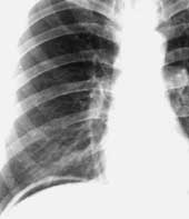
| Figure 1. Chest radiograph of patient with ahalasia. Dilated
oesophagus widen mediastinum to the right. |
|
|
|
| Figure 2. PA chest radiograph in case of oesophageal perforation
shows linear gas collection along right mediastinum border (arrows). |
|
|
|
| Figure 3. Patient with diaphragm oesophageal orifice hernia.
(a) PA chest radiograph shows additional shadow in right cardio-diaphragmatic
angle. |
|
|
|
| Figure 3. (b) Right lateral chest radiograph at the same
patient confirms presence of hernia in posterior mediastinum. |
|
|
|
| Figure 4. Diaphragmatic neoplasias and cysts (arrow) are seen
as additional shadow on the background of the lung which is not separated
from diaphragm on polypositional study. (a) PA chest radiograph. |
|
|
|
| Figure 4. (b) Left lateral chest radiograph. |
|
|
|
| Figure 5. Pneumoperitoneum – linear gas collection under right
diaphragm dome – after duodenum ulcer perforation. |
|
|
|
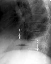
Figure 6. Left lateral chest radiograph shows gas-fluid level
under right diaphragm dome as abscess sign.
1 – right diaphragm dome,
2 – left diaphragm dome. |
|