Citation: News of Beam Diagnostics Belarus 2001 1-2: 4-10
Diagnostic imaging in peripheral lung cancer.
Golub G. D., Serova T. N.
Scientific Research Institute of Oncology and Medical Radiology named
for N. N. Aleksandrov, Minsk.
|
|
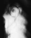
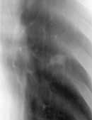
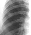
| Figure 1. Peripheral lung cancer simulating thin-wall cyst.
Aspergillum seen in the center of the tumor. |
|
|
|
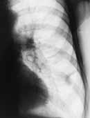
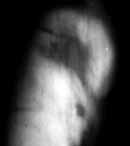
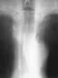
| Figure 2. Radiograph of the left lung. Peripheral lung cancer
developed on the background of the scar seen. |
|
|
|
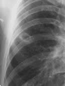
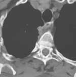
| Figure 3. Digital tomogram. Peripheral lung cancer (squamous)
presented as inhomogeneous nodule with speculated contour. |
|
|
|
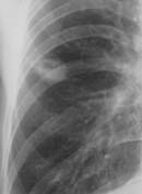
| Figure 4. Peripheral lung cancer (adenocarcinoma). Intensive
homogeneous shadow with strict, bumpy, speculated contour and tag to pleura
may be seen. |
|
|
|
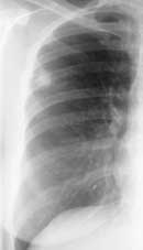
| Figure 5. Early lung cancer. Shadow with irregular contour and
central cavity seen. |
|
|
|
| Figure 6. Peripheral lung cancer. (а) Elongated shadow
looking like fibrous tag seen. |
|
|
|
| Figure 6. Peripheral lung cancer. (b) In 2 years tumour
considerably enlarged. Pleural tags and "path" to mediastinum seen. |
|
|
|
| Figure 7. Early lung cancer with cavitation. (а) Small
cavity at the level of the 6th rib. |
|
|
|
| Figure 7. Early lung cancer with cavitation. (b) In 6
months cavity considerably enlarged. |
|
|
|
| Figure 7. Early lung cancer with cavitation. (c) In 9
months tumour continues to enlarge. Partial cavity feeling may be seen. |
|
|
|
| Figure 8. Right lung radiograph shows superposition of peripheral
tumour shadows in S4 with 3rd rib. Lung nodule may be missed in sach a
case. |
|
|
|
| Figure 9. Cortical subpleural cancer. (а) Tomogram shows
small additional shadow at the right paratraheal region at the level of
2-3rd vertebrae. |
|
|
|
| Figure 9. Cortical subpleural cancer. (b) CT scan shows
bumpy tumour adjacent to vertebra body invading posterior mediastinum. |
|
|
|
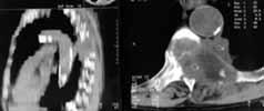
| Figure 10. Cortical subpleural cancer. CT scan and sagittal
reformat show paravertebral tumour with rib destruction. |
|