Citation: News of Beam Diagnostics Belarus 1998 4: 13-15.
Langergance-cell histiocytosis diagnostics.
Lazjuk I. I.1, Borisevich G. A.1, Sergeeva
A. A.1, Ratner T. P.2
1Belarussian Medical Academy of Postgraduate Education, 2Republican
Scientific Clinical Center of Pediatric Oncology and Hematology, Minsk.
|
|
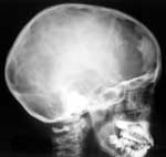
| Figure 1. Skull involvement in Langergance-cell histiocytosis.
Lateral skull radiograph shows geographic zone of destruction in frontal
bone. |
|
|
|
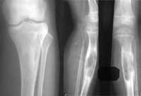
| Figure 2. Tibia involvement in Langergance-cell histiocytosis.
Lytic zones have a strict lobulated contours, and homogeneous structure. |
|
|
|
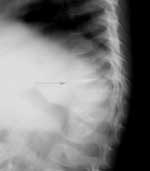
| Figure 3. Spine involvement in Langergance-cell histiocytosis. |
|
|
|
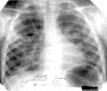
| Figure 4. PA chest radiograph in patient with Langergance-cell
histiocytosis shows redundancy and deformity of interstitial elements.
Big right lung cyst often complicated with suppuration may be seen. |
|