Citation: News of Beam Diagnostics Belarus 1998 4: 26-28.
CT and MRI diagnostics of head trauma sequela.
Bulaev I. V., Korytko S. S.
Scientific Research Institute of Radiation Medicine Clinics, Minsk.
|
|
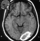
| Figure 1. Acute brain parenchymal haematoma. (a) Т1w
MR image. |
|
|
|
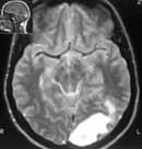
| Figure 1. Acute brain parenchymal haematoma. (b) Т2w. |
|
|
|
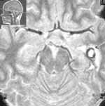
| Figure 2. Subacute haematoma of the left temporal lobe (arrow)
presents on T2w image as region with hyperintense centre (methaemoglobin)
and hypointense rim (haemosiderin). |
|